
What is a Vein Occlusion?
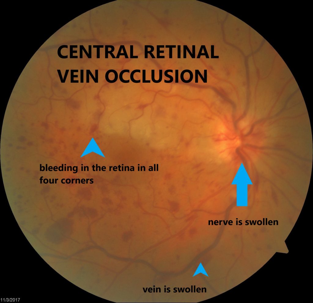
There are 2 different types of vein occlusions. Central Retinal Vein Occlusion (CRVO) and Branch Retinal Vein Occlusion (BRVO). The central retinal vein is a large main vein that carries blood away from the retina. If this vein partially or completely becomes blocked (occluded) it is called a CRVO. Off of the central vein are smaller branch veins that also carry blood away from the retina. If one of these smaller branch veins become partially or completely blocked (occluded) it is called a BRVO. Both of these veins are located in the retina which is the back, inner layer of the eye that converts light images to nerve signals and sends them to the brain. Having a central retinal vein occlusion is generally worse than having a branch retinal vein occlusion.
What are the symptoms of a vein occlusion?
In the early stages of vein occlusions, there are no symptoms. If swelling occurs in the macula, which is the area in the retina responsible for detailed vision, patients may begin to notice the blurry or distorted vision. There is usually no pain or physical sensations related to vein occlusions. Rarely, patients will present with new eye floaters from bleeding.
What causes central vein occlusion?
The cause of retinal vein occlusion is often hard to identify. Many times, blood pressure is the culprit. Most retinal specialists think there is an underlying anatomic reason that puts some patients at risk for vein occlusion. Blockage of smaller veins (BRVO) in the retina often occurs in places where retinal arteries that have been thickened or hardened by artery disease cross over and place pressure on a retinal vein.
Risk factors
- Diabetes
- Vein occlusion in one eye in the past
- Hypertension
- Age over 55 years old
- High eye pressure, glaucoma
- Blood clots
- Blood cancer
- Cardiovascular disease
- High Body Mass Index (BMI)
What is the treatment for branch retinal vein occlusion?
Many people can regain useful vision with treatment. However, vision rarely returns to normal. There is no way to reverse or open the blockage. Retina specialists primarily treat the swelling that occurs in the retinal tissue related to the blocked vein. This swelling is called macular edema. Rarely, the blood flow can be so poor after a vein occlusion that the small, abnormal blood vessels may form that require laser or eye injection therapy (neovascularization). You may need treatment to prevent another blockage from forming in the same eye or to reduce the damage from the original occlusion.
How serious is a vein occlusion?
People with vein occlusions often regain useful vision. The final visual outcome can vary depending on the degree of blood flow and swelling in the retina.
What can I do to prevent a vein occlusion?
Retinal vein occlusion is a sign of a general blood vessel (vascular) disease. Maintaining a healthy lifestyle is important. Taking certain measures such as a low-fat diet, regular exercise, keeping your weight down, controlling diabetes and not smoking can be very helpful in preventing retinal vein
occlusions. If an occlusion has effected one eye, you may want to speak to your medical doctor about taking steps to reduce the chances of having another vein occlusion in your other eye.