
Epiretinal Membrane / Macular Pucker
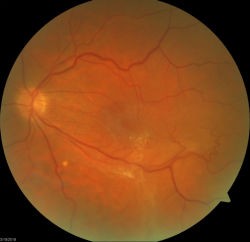 The back of the eye is called the retina. In the center of the retina is the macula. The macula is the part of the eye that gives us our most detailed central vision. Sometimes, a thin, fibrous membrane or “scar tissue can form over the macula. This scar tissue can cause distortion in one’s central vision. This is called an EPIRETINAL MEMBRANE or MACULAR PUCKER. Other names like Cellophane Retinopathy or Pre Retinal Fibrosis are also sometimes used to describe the same condition. You may see it abbreviated as ERM on your eye doctor’s notes.
The most common cause of a macular pucker or epiretinal membrane is simply getting older. The back of the eye is filled with a thick jelly-like substance called the vitreous. When the vitreous strips away from the retina sometimes it liberates normal cells into the middle part of the eye. These cells often settle over the center vision and grow slowly as a sheet over time. This sheet of scar tissue will begin to cause distortion in the detailed vision. Other causes of a macular pucker can include inflammatory diseases, trauma, bleeding, or can sometimes with no good reason at all.
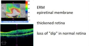
This can be diagnosed easily by having a dilated eye examination and special high definition photographs taken of the retina.
 In many cases, no treatment is needed for an epiretinal membrane as it is not dangerous to the health of the eye. If the scar tissue worsens and causes enough disturbance in vision, then a surgical procedure can be performed by a retina surgeon. The procedure is called an epiretinal membrane peel. The thin fibrous membrane is surgically removed off of the macula and at the same time, the vitreous jelly is removed from the back of the eye and replaced with a saline solution. This is called a vitrectomy.
The recovery after an epiretinal membrane peel and vitrectomy is fairly simple. A series of antibiotic and anti-inflammatory eye drops may be used for several weeks and postoperative visits with your retinal specialist will be necessary to observe the improvement in vision. No face down positioning is usually required. Most patients will have a significant improvement in vision and distortion after surgery.